

Top 10 Friday Beautiful Sciences of 2007
Everyone is making lists for 2007, and I thought I would too. Here's a list of my favourite Friday beautiful science posts of 2007. In particular, I'd like to take issue with Phil Plait's (of Bad Astronomy) statement:
Astronomy is arguably the most beautiful of the sciences. I’m biased, of course, but it’s nearly impossible to gaze upon a picture of a galaxy, a moon, a nebula, and not see in it something compellingly artistic. Sometimes it’s the color, sometimes the shape, and sometimes it’s the knowledge that we can understand the subject of the picture itself.Many of the pictures I present are from biology, also a beautiful science. In no particular order, I present my top ten picks of Friday beautiful science:

Today's Friday Beautiful Science is a depiction of the tree of life, done as a sphere. Previously, I've discussed the limitations of depicting phylogeny on a standard tree, and suggested that doing it as a circle or a sphere would be more useful. Turns out I'm not the only one who thinks that. This is one particular 3-D visualization that I've taken from Tim Hughes at the University of Bergen (he has several others at his site). Yet other 2-dimensional visualizations can bee seen here.
-----
 This weeks Friday beautiful science comes from Eye of Science:
 Scales from the skin of a shark. These sharply pointed placoid scales are also known as dermal teeth or denticles. They give the shark's skin the feel of sandpaper. The tip of each scale is made of dentine overlayed with dental enamel. The lower part of each scale, which anchors it into the skin, is made of bone. The scales disrupt turbulence over the skin, considerably reducing the drag on the shark as it swims. This design has been investigated by engineers for use on the surfaces of aircraft and boats. Coloured scanning electron micrograph, Magnification: x70.Eye of Science is a:
two-person team of photographer and biologist,
our aim is to combine scientific exactness with aesthetic
appearances, and thereby help to bridge the gap
between the world of science and the world of art.
-----
Today's Friday beautiful science comes from the lab of Julie Theriot. These are sped up movies of Listeria bacteria inside human cells. Listeria actually attaches to the actin filaments inside the cell, and uses the actin to force itself around through the cell (and to force its way into neighboring cells). On the left side of the human cell, you can see some bacteria stretching the cytoplasm. If there had been another human cell adjacent to this one, the Listeria cell likely would have punctured the membrane of the human cell, and moved directly into the adjacent human cell. Super cool.
From the Theriot lab website:
Listeria monocytogenes is a small rod shaped gram-positive bacterium that is ubiquitous in the environment, in the soil, on plants and animals. Listeriosis (the state of Listeria infection) is associated with eating of unpasteurized cheese or dairy products, or consumption of contaminated vegetables. Infection occurs primarily in newborns and infants, elderly or immunocompromised individuals, or pregnant women (mother is asymptomatic or has influenza-like syndrome, but the newborn can acquire it during birth, or infection can cause abortion or premature delivery). According to the CDC about 1500 cases are reported in the United States each year, mostly in the aforementioned high risk groups.Check out this part of Dr. Theriot's site for more cool movies.
Listeria enters the host along with infected food. Most of the bacteria will be killed by acid in the stomach, but the surviving bacteria invade the cells of the intestinal tract, going from cell to cell and thereby spreading the infection laterally. Intracellular movement of the bacteria is essential for this lateral infection to occur, and Listeria has been described to move with a "comet-tail" or like an "actin rocket". The Theriot Laboratory is studying how Listeria moves within cells, and results of this research may someday be used to generate vaccines or other means to prevent infection by Listeria. Currently, the only preventive measures are thorough cooking and cleaning of food, as well as pasteurization of dairy products. Treatment of Listeriosis primarly depends on antibiotics.
-----
 This Friday's beautiful science is a photo of the surface of Europa taken by the Galileo probe in 1998. The American Museum of Natural History in New York is having a show of fantastic photos taken from various space probes. Michael Benson put the photographs together using raw data from NASA (many of the shots are composites). If you can get to New York, see the show (and please, comment on it here).
For those of us unable to make it to the show, Michael Benson has put together a book. I got it last week, it's fantastic. The photos are all stunning. The very few photos that I've seen that are better than the photos in his book are the very recently taken photos of the sun taken by Hinode, and of Saturn from Cassini.
Check out the book here:
-----

This is an image taken of a siphonophore taken from the Arctic in 2005. Stunning.
-----
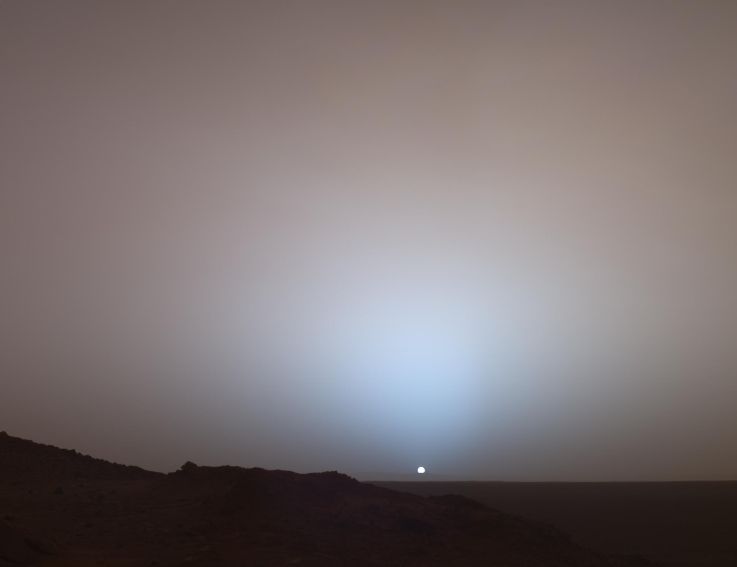
Here's an old shot (2005) from the Mars rover Spirit. It's photos like this why I got involved in science.
-----
 Today's Friday beautiful science comes from the book Evolution by Jean-Baptiste De Panafieu. As described:
 Each chapter is made up of a short text that illuminates one theme of the evolutionary process-repetition, adaptation, polymorphism, sexual selection-and a series of exquisitely composed photographs of skeletons against a black background. Approximately three hundred photographs of whole skeletons or details have been made possible by the French National Museum of Natural History. The reader learns, by experiencing each text and photograph together, how the structure of every creature has been shaped by its environmental and genetic inheritance.This one's on my Christmas list.
More photos below.


-----

via Bad Astronomy, I bring you Iapetus. Again! Blow this image up, turn out the lights, and gaze at it on your screen. It's beautiful. (These images were taken by Cassini on Sep 10, 2007 - so they're really very new).
Update: Even more amazingness from Cassini:

-----
 Today's Friday beautiful science comes from the AKARI satellite, launched by the Japanese Space Agency. From their website:
 The infrared astronomy satellite AKARI started the regular observations in May 2006. In the last one year, AKARI has carried out the All-Sky Survey observations to map the entire sky, as well as thousands of pointed observations of selected targets. Here we show the beautiful images constructed from the AKARI All-Sky Survey data; The entire sky in the mid-infrared light, the far-infrared image of the constellation Orion and the Milky Way, and the far-infrared image of the Cygnus-X region.This photo is a composite of many thousands of images. The line running through the middle of the photo is the Milky Way.
Sadly, the satellite is nearly done its mission. It was designed to last for 550 days, and has nearly run out of liquid helium (which helps keep its sensors cool).
I'm pretty excited about this photo, and have already had a print made which I'll frame this weekend and hang in my living room. I hope you like it as much as I do.
-----
Here's a video I've been dying to post since I first saw it a few months ago.
As described on the Cornell Mushroom Blog:
Here’s something I know you’ve all been dying to see. A video of one of the most compellingly jaw-dropping spectacles in mycology, condensed from four days of electrifying footage. What you can’t see is the stink, the awesome stink associated with this event. It caused our noble departmental photographer, Kent Loeffler, to vent the noxious, carrion-like fumes into the fourth floor hallway outside my office. We have all suffered here in the service of science.More cool time-lapse videos here.
There are two fungi in this video. On your right, the common pinkish-stemmed Phallus ravenelii. On your left, the rarer, paler, netted stinkhorn, Dictyophora duplicata. Note also the guest appearance by Hubert the fly (aka “The Vector”) late in the video! Oh! the humanity.
To preemptively answer your insightful questions, let me clarify a few points. The stinkiness is part of the dispersal mechanism of the crafty stinkhorn. The green goop covering the heads is a spore slurry that stinks in a sultry way and attracts flies and such. Insects disperse the spores on their little feet. Why does the mushroom look this way? I can’t say, but I can assure you that you’re not the first to notice a certain resemblance. The stinkhorns belong to an order of fungi called the Phallales. They have been causing trouble for a long time, and first got their suggestive Latin name in 1564.
via Small Things Considered.


3 comments:
I love your blog....and this post is amazing....and yes I am adding you to my blogroll..
( btw is your avatar Smoking Man from Xfiles ? )
please come see me over at Watergate Summer....
Thanks. Yes, that is the smoking man.
Happy New Year.
-F.
wow.. amazing post... thank you
I am adding you to my daily read just on this post alone...
constantskeptic.com
Post a Comment