Showing posts sorted by relevance for query friday beautiful science. Sort by date Show all posts

Friday, December 28, 2007

Top 10 Friday Beautiful Sciences of 2007

Everyone is making lists for 2007, and I thought I would too. Here's a list of my favourite Friday beautiful science posts of 2007. In particular, I'd like to take issue with Phil Plait's (of Bad Astronomy) statement:

Astronomy is arguably the most beautiful of the sciences. I’m biased, of course, but it’s nearly impossible to gaze upon a picture of a galaxy, a moon, a nebula, and not see in it something compellingly artistic. Sometimes it’s the color, sometimes the shape, and sometimes it’s the knowledge that we can understand the subject of the picture itself.
Many of the pictures I present are from biology, also a beautiful science. In no particular order, I present my top ten picks of Friday beautiful science:


Today's Friday Beautiful Science is a depiction of the tree of life, done as a sphere. Previously, I've discussed the limitations of depicting phylogeny on a standard tree, and suggested that doing it as a circle or a sphere would be more useful. Turns out I'm not the only one who thinks that. This is one particular 3-D visualization that I've taken from Tim Hughes at the University of Bergen (he has several others at his site). Yet other 2-dimensional visualizations can bee seen here.

-----

This weeks Friday beautiful science comes from Eye of Science:
Scales from the skin of a shark. These sharply pointed placoid scales are also known as dermal teeth or denticles. They give the shark's skin the feel of sandpaper. The tip of each scale is made of dentine overlayed with dental enamel. The lower part of each scale, which anchors it into the skin, is made of bone. The scales disrupt turbulence over the skin, considerably reducing the drag on the shark as it swims. This design has been investigated by engineers for use on the surfaces of aircraft and boats. Coloured scanning electron micrograph, Magnification: x70.
Eye of Science is a:
two-person team of photographer and biologist,
our aim is to combine scientific exactness with aesthetic
appearances, and thereby help to bridge the gap
between the world of science and the world of art.

-----


Today's Friday beautiful science comes from the lab of Julie Theriot. These are sped up movies of Listeria bacteria inside human cells. Listeria actually attaches to the actin filaments inside the cell, and uses the actin to force itself around through the cell (and to force its way into neighboring cells). On the left side of the human cell, you can see some bacteria stretching the cytoplasm. If there had been another human cell adjacent to this one, the Listeria cell likely would have punctured the membrane of the human cell, and moved directly into the adjacent human cell. Super cool.

From the Theriot lab website:
Listeria monocytogenes is a small rod shaped gram-positive bacterium that is ubiquitous in the environment, in the soil, on plants and animals. Listeriosis (the state of Listeria infection) is associated with eating of unpasteurized cheese or dairy products, or consumption of contaminated vegetables. Infection occurs primarily in newborns and infants, elderly or immunocompromised individuals, or pregnant women (mother is asymptomatic or has influenza-like syndrome, but the newborn can acquire it during birth, or infection can cause abortion or premature delivery). According to the CDC about 1500 cases are reported in the United States each year, mostly in the aforementioned high risk groups.

Listeria enters the host along with infected food. Most of the bacteria will be killed by acid in the stomach, but the surviving bacteria invade the cells of the intestinal tract, going from cell to cell and thereby spreading the infection laterally. Intracellular movement of the bacteria is essential for this lateral infection to occur, and Listeria has been described to move with a "comet-tail" or like an "actin rocket". The Theriot Laboratory is studying how Listeria moves within cells, and results of this research may someday be used to generate vaccines or other means to prevent infection by Listeria. Currently, the only preventive measures are thorough cooking and cleaning of food, as well as pasteurization of dairy products. Treatment of Listeriosis primarly depends on antibiotics.
Check out this part of Dr. Theriot's site for more cool movies.

-----

This Friday's beautiful science is a photo of the surface of Europa taken by the Galileo probe in 1998. The American Museum of Natural History in New York is having a show of fantastic photos taken from various space probes. Michael Benson put the photographs together using raw data from NASA (many of the shots are composites). If you can get to New York, see the show (and please, comment on it here).

For those of us unable to make it to the show, Michael Benson has put together a book. I got it last week, it's fantastic. The photos are all stunning. The very few photos that I've seen that are better than the photos in his book are the very recently taken photos of the sun taken by Hinode, and of Saturn from Cassini.

Check out the book here:


-----


This is an image taken of a siphonophore taken from the Arctic in 2005. Stunning.

-----
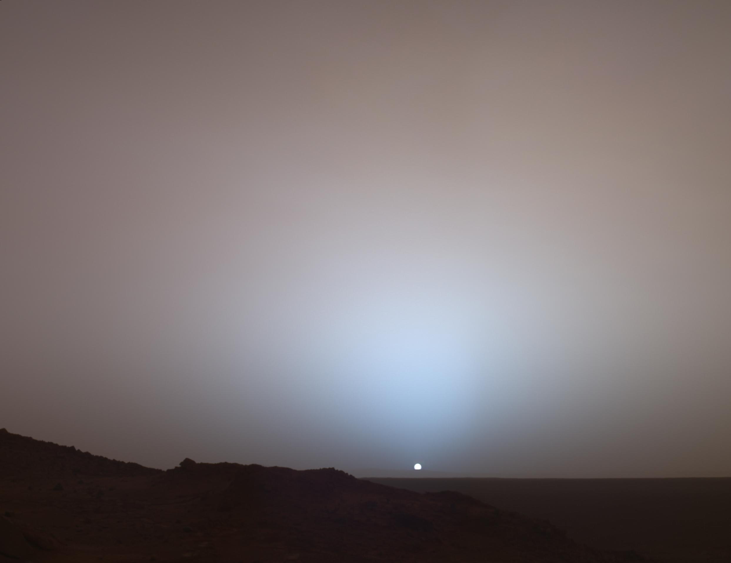

Here's an old shot (2005) from the Mars rover Spirit. It's photos like this why I got involved in science.

-----

Today's Friday beautiful science comes from the book Evolution by Jean-Baptiste De Panafieu. As described:
Each chapter is made up of a short text that illuminates one theme of the evolutionary process-repetition, adaptation, polymorphism, sexual selection-and a series of exquisitely composed photographs of skeletons against a black background. Approximately three hundred photographs of whole skeletons or details have been made possible by the French National Museum of Natural History. The reader learns, by experiencing each text and photograph together, how the structure of every creature has been shaped by its environmental and genetic inheritance.
This one's on my Christmas list.


More photos below.


-----


via Bad Astronomy, I bring you Iapetus. Again! Blow this image up, turn out the lights, and gaze at it on your screen. It's beautiful. (These images were taken by Cassini on Sep 10, 2007 - so they're really very new).

Update: Even more amazingness from Cassini:


-----

Today's Friday beautiful science comes from the AKARI satellite, launched by the Japanese Space Agency. From their website:
The infrared astronomy satellite AKARI started the regular observations in May 2006. In the last one year, AKARI has carried out the All-Sky Survey observations to map the entire sky, as well as thousands of pointed observations of selected targets. Here we show the beautiful images constructed from the AKARI All-Sky Survey data; The entire sky in the mid-infrared light, the far-infrared image of the constellation Orion and the Milky Way, and the far-infrared image of the Cygnus-X region.
This photo is a composite of many thousands of images. The line running through the middle of the photo is the Milky Way.

Sadly, the satellite is nearly done its mission. It was designed to last for 550 days, and has nearly run out of liquid helium (which helps keep its sensors cool).

I'm pretty excited about this photo, and have already had a print made which I'll frame this weekend and hang in my living room. I hope you like it as much as I do.

-----


Here's a video I've been dying to post since I first saw it a few months ago. Blogger now makes it easy to post videos that aren't on YouTube. The video function choked today, and I had to get put the video on Youtube.

As described on the Cornell Mushroom Blog:
Here’s something I know you’ve all been dying to see. A video of one of the most compellingly jaw-dropping spectacles in mycology, condensed from four days of electrifying footage. What you can’t see is the stink, the awesome stink associated with this event. It caused our noble departmental photographer, Kent Loeffler, to vent the noxious, carrion-like fumes into the fourth floor hallway outside my office. We have all suffered here in the service of science.

There are two fungi in this video. On your right, the common pinkish-stemmed Phallus ravenelii. On your left, the rarer, paler, netted stinkhorn, Dictyophora duplicata. Note also the guest appearance by Hubert the fly (aka “The Vector”) late in the video! Oh! the humanity.

To preemptively answer your insightful questions, let me clarify a few points. The stinkiness is part of the dispersal mechanism of the crafty stinkhorn. The green goop covering the heads is a spore slurry that stinks in a sultry way and attracts flies and such. Insects disperse the spores on their little feet. Why does the mushroom look this way? I can’t say, but I can assure you that you’re not the first to notice a certain resemblance. The stinkhorns belong to an order of fungi called the Phallales. They have been causing trouble for a long time, and first got their suggestive Latin name in 1564.
More cool time-lapse videos here.

via Small Things Considered.
Digg!

Friday, March 7, 2008

Friday beautiful science

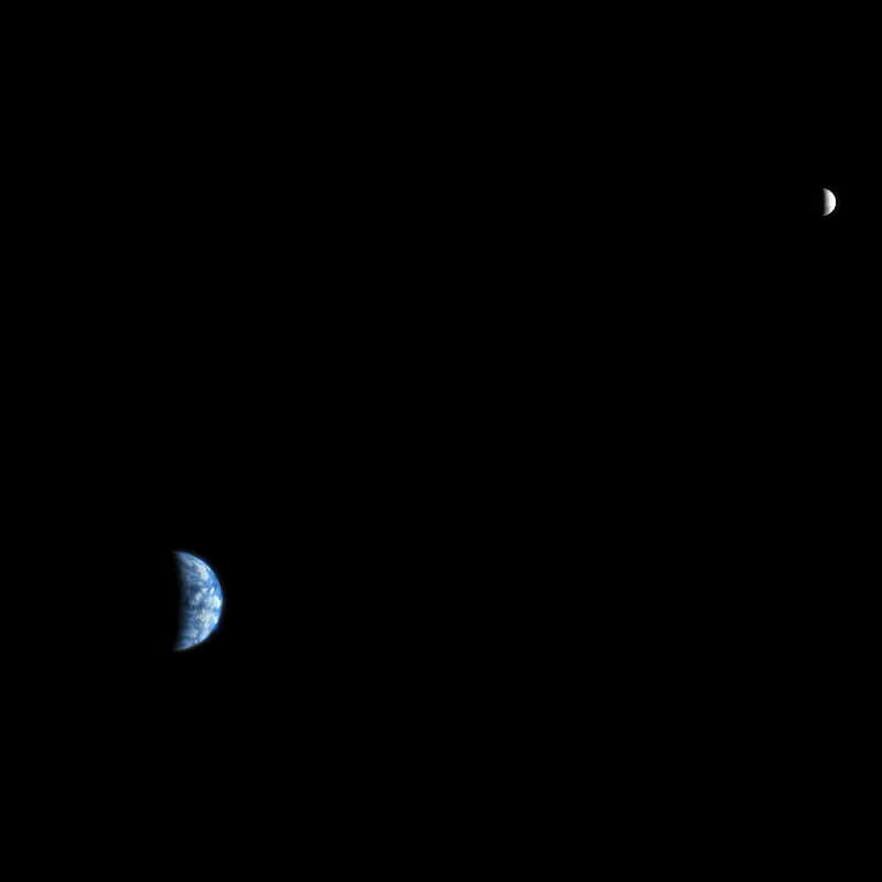
Today's Friday beautiful science is us. Here's a shot of our tiny little planet, as seen from Mars. The High Resolution Imaging Science Experiment (HIRISE) is orbiting Mars:

Launched in August 2005, the High Resolution Imaging Science Experiment (HiRISE) is flying onboard the Mars Reconnaissance Orbiter (MRO) mission. HiRISE will investigate deposits and landforms resulting from geologic and climatic processes and assist in the evaluation of candidate landing sites.
But in this particular image, they've pointed their cameras at earth to get a shot of the earth and the moon.

As an aside, I wanted to put this shot down as the Friday beautiful science this week, but Mrs. Factician thinks it's too difficult to look at. I'll let you be the judge.


This is another shot taken by the HIRISE. It's catching an avalanche in real time. The top of the mountain is on the left side of the photo, sloping downwards to the right. Those clouds to the right are dust thrown up by the avalanche. Absolutely beautiful. This photo is false colored.
Material, likely including fine-grained ice and dust and possibly including large blocks, has detached from a towering cliff and cascaded to the gentler slopes below. The occurrence of the avalanches is spectacularly revealed by the accompanying clouds of fine material that continue to settle out of the air. The largest cloud (upper images) traces the path of the debris as it fell down the slope, hit the lower slope, and continues downhill, forming a billowing cloud front. This cloud is about 180 meters (590 feet) across and extends about 190 m (625 ft) from the base of the steep cliff. Shadows to the lower left of each cloud illustrate further that these are three dimensional features hanging in the air in front of the cliff face, and not markings on the ground (sun is from the upper right).
Wow.

Digg!

Friday, May 16, 2008

Friday beautiful science

Sorry I've been quiet for a while. Various personal issues have kept me fairly occupied (including flying out to California for a job interview at a biotech company).

Today's Friday beautiful science is something I've lusted after ever since I first saw it. It's a 3-dimensional map of our best data about the Milky Way galaxy, mapped out in painstaking detail inside of a cube of glass. It's simply beautiful.

Behold: a galaxy suspended in a glass cube.

A laser was used to etch around 80,000 of the stars in the Milky Way, using three-dimensional data from the Japan’s National Astronomical Observatory.

Production of the cube was motivated by the urge to see a galaxy in three dimensions. In encyclopedias and such, galaxies can only be viewed in two: we wanted to do so from all directions. Even the Magellan Cloud, comparatively close to us, is outside this cube. Space can be sparsely filled, one could say…or rather, there are great extremes in density.
I've made prints of many of my other Friday beautiful science photos. Perhaps some day I'll save enough Japanese yen to be able to purchase this little beauty...

Digg!

Friday, June 13, 2008

Friday beautiful science

Today's Friday beautiful science comes a bit late.  It's a picture of the Phoenix lander, landing on Mars.  Check it out:  

This amazing image was captured as Phoenix came in for its Mars landing on May 25, 2008. The HiRISE camera on Mars Reconnaissance Orbiter pointed at Phoenix, which is seen here against the background of a 10-kilometer-diameter crater called Heimdall. The dramatic view makes it appear that Phoenix is falling into the crater, but in fact Phoenix was 20 kilometers closer to HiRISE than Heimdall, and it landed nowhere near the crater. The photo was taken 20 seconds after Phoenix' parachute opened.


Posts should become more frequent again. I'm preparing to move to California, but some of the most brutal parts are behind me (I hope) and I should be able to at least keep a regular Friday beautiful science coming.

Digg!

Friday, June 29, 2007

Friday beautiful science

This weeks Friday beautiful science comes from Eye of Science:

Scales from the skin of a shark. These sharply pointed placoid scales are also known as dermal teeth or denticles. They give the shark's skin the feel of sandpaper. The tip of each scale is made of dentine overlayed with dental enamel. The lower part of each scale, which anchors it into the skin, is made of bone. The scales disrupt turbulence over the skin, considerably reducing the drag on the shark as it swims. This design has been investigated by engineers for use on the surfaces of aircraft and boats. Coloured scanning electron micrograph, Magnification: x70.
Eye of Science is a:
two-person team of photographer and biologist,
our aim is to combine scientific exactness with aesthetic
appearances, and thereby help to bridge the gap
between the world of science and the world of art.

Digg!

Friday, June 15, 2007

Friday beautiful science


Today's Friday beautiful science focuses on the work of Felice Frankel. She's a photographer at MIT who works with scientists to take cool photos of their work. The photo above was taken of a ferromagnetic fluid on a glass slide with seven magnets underneath. There are several other of her photos displayed in a recent New York Times profile of her. She has one book of photos (On the Surface of Things) that's out of print, and another (Envisioning Science: The Design and Craft of the Science Image) that is intended for photographers.

If anyone can point me towards a source for her prints, I would appreciate it.

Digg!

Friday, September 28, 2007

Friday beautiful science


Today's Friday beautiful science comes from the National Science Foundation's Visualization Challenge. This photo was one of two co-winners for the 2007 prize. (I have featured a previous prizewinner here). Though this looks like a dragon running head on towards you, it is in fact a CT scan of the underside of a woman's head, highlighting the sinuses:

Human anatomy it may be, but the airways that riddle the space behind our noses take on an alien aspect in this unearthly rendering created by Kai-hung Fung, a radiologist at the Pamela Youde Nethersole Eastern Hospital in Hong Kong. A computed tomography (CT) scan from a 33-year-old Chinese woman being examined for thyroid disease provided the raw data for Fung's rendering. He stacked together 182 thin CT "slices" to create a 3D image looking upward at the sinuses from underneath the head. Normally, CT renderings meld slices together into smooth surfaces, but, in what he terms the "Rainbow Technique," Fung instead broke them apart, creating a topographical map of the airspaces described by the contour lines of individual slices, and colored according to the density of the tissues that border them.
You can see all the winners here.

Digg!

Friday, October 19, 2007

Friday beautiful science

Today's Friday beautiful science comes from the Nanotechnology Now art gallery at the University of Cambridge Department of Materials Science and Metallurgy:

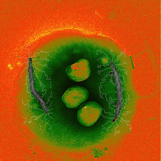
The image shows the magnetic field lines in a single bacterial cell. The fine white lines are the magnetic field lines in the cell, which were measured using off-axis electron holography. Such bacteria live in sediments and bodies of water, and move parallel to geomagnetic field lines as a result of the torque exerted on their magnetosome chains by the earth's magnetic field.

Digg!

Friday, April 11, 2008

Friday beautiful science

Today's Friday Beautiful Science is a photo of the moon Phobos. Phobos orbits Mars, and is orders of magnitude smaller than Earth's moon (compare Earth's moon at 3400 km in diameter to Phobos at 19-21 km in diameter). As you can clearly see in the photo, Phobos is not regularly shaped. From the Planetary Science blog:

Mars Reconnaissance Orbiter approached to within 6,800 kilometers (4,200 miles) of Phobos to capture this enhanced-color view of the Martian moon on March 23, 2008. The color is from infrared, red, and blue-green channels on the camera, so it represents light shifted slightly longer in wavelength than human eues can see, which emphasizes subtle colorations on the moon. The color view shows that the material surrounding the giant crater Stickney (on the left side of the moon) appears gray while the rest of the moon appears reddish. The grayer material is likely fresher material.
Click on the photo to get a large majestic view of Phobos.

Digg!

Friday, October 26, 2007

Friday beautiful science


I'm recycling an old image for today's Friday Beautiful Science. I'm on vacation, and am spending *very* little of it online. It's a shot of a sunset taken from one of the Mars Rovers. I'm currently enjoying the sunrises and sunsets in the mountains of Quebec, Canada. A little less desolate than Mars, but just as beautiful.

Back next week...

Digg!

Wednesday, August 15, 2007

Friday beautiful science - On Wednesday!


I missed last week's Friday beautiful science. Between doctor's appointments and being strung out on prescription medication, I couldn't even get my s*#t together enough to post a photo. So I'm doing two this week. One today, and one on Friday. I found today's at Bad Astronomy. This is a photo of the star Mira, found 400 light years from Earth. Here's Phil's description:

Mira is definitely wonderful, in the sense of evoking wonder. And now we have found out it’s even more amazing than we thought. Most stars near the Sun orbit the center of the Galaxy at roughly the same speed, but some are faster than others. Mira, it so happens, is plowing through this local region of space at about 130 kilometers per second (about 80 miles per second). There is gas and dust out there, a thin haze floating among the stars. As Mira screams through this fog, the gas it is ejecting as it convulses is blown backwards, leaving a long tail behind it — imagine running down the street with a smoke bomb in your hand and you’ll get the idea.

Now take another look at the image at the top of this page. Mira is on the right hand side, and is moving left to right. The long tail of ejected material is incredible — it’s 13 light years long! It has taken Mira 30,000 years to move this distance, which means that the material in the left hand side of the tail was ejected 30 millennia ago. If you look at the location of the star itself, you’ll see a parabolic arc in front of it; that’s the bow shock, where Mira’s ejected material is slamming into the material between stars (called the interstellar medium or ISM).

He also describes how the material for Mira will be involved in creating new solar systems. Very cool stuff, head over there and read it. Now.

For full resolution photo, go here.

NASA includes a timeline on the age of Mira's tail:

Digg!

Friday, November 16, 2007

Friday beautiful science

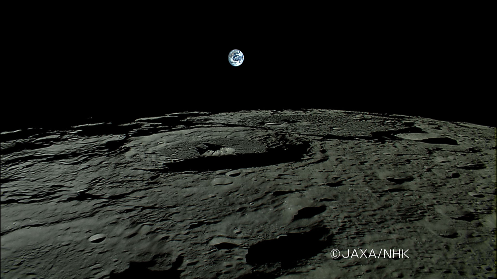
For the second week in a row, the Friday beautiful science comes from the Japanese moon orbiter Kaguya/Selene. They caught this beautiful photo of the earth rising over the horizon of the moon. Very pretty. They also have a short movie on their site that they've shown in HD on TV programs in Japan and Canada (hopefully soon here in the U.S. as well).

I confess, I'm a tiny bit disappointed with the Japanese Space Agency, however. Unlike NASA, they don't seem to release full resolution photos. Just these low to medium resolution jpegs, marked with the copyright. It's a pity, because I'm just nerd enough to print these types of photos, frame them and hang them in my house, and this photo is barely high enough resolution to make a decent sized print. I think NASA makes a great choice by distributing high resolution photos as a public relations tool to get folks excited about their work. JAXA should do the same.

Digg!

Thursday, May 17, 2007

Friday beautiful science

Today's Friday beautiful science is an animation created by the animation studio XVIVO using funding from Howard Hughes Medical Institute and Harvard University. It's a fantastic illustration of all the things that need to go inside a white blood cell during its survey of a blood vessel and subsequent invasion into infected tissue. It's reasonably accurate, right down to the structural level of the proteins that are depicted. Though the material is probably suitable for mid to upper level biology undergrads, it's still beautiful to watch.


There's a slightly lighter version put to music, if you're not all that interested in the details (or, if you're a biologist, and you recognize everything going on there without the narration).


I confess, these two are so good they give me chills...

Digg!

Friday, April 18, 2008

Friday beautiful science


Today's Friday beautiful science is a shot of bacteria spreading across a petri plate, with false color added to make them more beautiful. This looks like a Bacillus mycoides type of growth, but I couldn't find the identity on the original website.

Go on over to Eshel Ben-Jacob's website, where there are all kinds of beautifully rendered shots of bacterial growth.

Digg!

Friday, August 3, 2007

Friday beautiful science

Today's Friday beautiful science comes from the AKARI satellite, launched by the Japanese Space Agency. From their website:

The infrared astronomy satellite AKARI started the regular observations in May 2006. In the last one year, AKARI has carried out the All-Sky Survey observations to map the entire sky, as well as thousands of pointed observations of selected targets. Here we show the beautiful images constructed from the AKARI All-Sky Survey data; The entire sky in the mid-infrared light, the far-infrared image of the constellation Orion and the Milky Way, and the far-infrared image of the Cygnus-X region.
This photo is a composite of many thousands of images. The line running through the middle of the photo is the Milky Way.

Sadly, the satellite is nearly done its mission. It was designed to last for 550 days, and has nearly run out of liquid helium (which helps keep its sensors cool).

I'm pretty excited about this photo, and have already had a print made which I'll frame this weekend and hang in my living room. I hope you like it as much as I do.

Digg!

Friday, February 29, 2008

Friday beautiful science

Today's Friday beautiful science is a shot of a fruitfly (Drosophila melanogaster) larva trapped in a water droplet. From the Nature website:

This image of a live Drosophila larva in a water droplet has won the photographic competition that forms part of celebrations marking the 200th anniversary of the Royal Netherlands Academy of Arts and Sciences (KNAW). Winner Robert Markus, of the Biological Research Center of the Hungarian Academy of Sciences in Szeged, received the award at a ceremony on 25 February in Amsterdam.

...

Markus took his photograph to show how blood cells affect the fruitfly's immune system. "By identifying blood-cell-specific genes, we can generate transgenic Drosophila strains in which the blood cells express green fluorescent protein, so that they are visualized in vivo, making in vivo research possible on the immune system," he explained.
If you know Dutch, head on over to the prize website, and check out more info.

Digg!

Friday, June 27, 2008

Friday beautiful science


Today's Friday beautiful science comes from The Vector Site. They have a nice primer for young students on how evolution by natural selection works. One of the photos they show is of the leafy sea dragon. They look like kelp, but they're sea dragons. Look again?

I've seen these in tanks at an aquarium before, and they are truly stunning to behold. As much as I hate to admit it, Wikipedia has a pretty good description of them here.

Digg!

Friday, March 28, 2008

Friday beautiful science

Today's Friday Beautiful Science comes from the University of Maryland Biotechnology Institute. They're genetically engineering mosquitoes, with the hopes of making a malaria resistant mosquito. See more pictures here.

Digg!

Friday, January 25, 2008

Friday beautiful science

Today's Friday beautiful science comes from the Mercury Messenger mission:

One week ago, the MESSENGER spacecraft transmitted to Earth the first high-resolution image of Mercury by a spacecraft in over 30 years, since the three Mercury flybys of Mariner 10 in 1974 and 1975. MESSENGER's Wide Angle Camera (WAC), part of the Mercury Dual Imaging System (MDIS), is equipped with 11 narrow-band color filters, in contrast to the two visible-light filters and one ultraviolet filter that were on Mariner 10's vidicon camera. By combining images taken through different filters in the visible and infrared, the MESSENGER data allow Mercury to be seen in a variety of high-resolution color views not previously possible. MESSENGER’s eyes can see far beyond the color range of the human eye, and the colors seen in the accompanying image are somewhat different from what a human would see.
Wow!

Digg!

Friday, July 20, 2007

Friday beautiful science

Today's Friday beautiful science is a hybrid photo of 4 photos taken by the Cassini space probe on New Year's Eve 2004. They show Saturn's moon Iapetus. This week, scientists at the Jet Propulsion Laboratory unveiled a model for how it is that Iapetus ended up with the ridge running across the moon:

"We've modeled how Iapetus formed its big, spin-generated bulge and why its rotation slowed down to its present nearly 80-day period. As an unexpected bonus, Iapetus also told us how old it was," said Dennis Matson, Cassini project scientist at JPL. "You would expect a very fast-spinning moon to have this bulge, but not a slow-spinning moon, because the bulge would have been much flatter."

Scientists calculate Iapetus originally rotated much faster -- at least five hours, but less than 16 hours per revolution. The fast spin gave the moon an oblate shape that increased the surface area (in the same way the surface area of a round balloon stretches when the balloon is pressed into an oblate shape). By the time the rotation slowed down to a period of 16 hours, the outer shell of the moon had frozen. Furthermore, the surface area of the cold moon was now smaller. The excess surface material was too rigid to go back smoothly into the moon. Instead, it piled up in a chain of mountains at the equator.

"Iapetus' development literally stopped in its tracks," said Castillo. "In order for tidal forces to slow Iapetus to its current spin rate, its interior had to be much warmer, close to the melting point for water ice." The challenge in developing a model of how Iapetus came to be "frozen in time" has been in deducing how it ever became warm enough to form a bulge in the first place, and figuring out what caused the heat source to turn off, leaving Iapetus to freeze.

The heat source had to have a limited life span, to allow the moon's crust to rapidly become cold and retain its immature shape. After looking at several models, scientists concluded that the heat came from its rocks, which contain short-lived radioactive isotopes aluminum-26 and iron-60 (which decay very rapidly on a geologic timescale). Since these elements decay at a known rate, this allowed scientists to "carbon date" Iapetus by using aluminum-26 instead of carbon. Scientists calculate the age of Iapetus to be roughly 4.564 billion years old.
Via Bad Astronomy.

Digg!